We reported magnetooptical properties of Tb3+ in single crystals of Tb3Fe5O12 and Tb3Ga5O12 for ion occupying sites of D2 symmetry in the garnets structure. It is shown that in the employed Voigt geometry the magnetic linear birefringence and the dichroism reach values 10-4, and have a strong dependence on the wavelength and a strong anisotropy. The absorption spectra were obtained at temperatures of 30K, 100K using magnetic field up to 25 kOe applied parallel and perpendiculare to the electric vector E linearly polarized light on the 7F6 – 7F0 and 7F6 – 7F1 optical transitions region. The aim of this research was revealing of a role of contributions of exchange interaction and a crystal field in splitting of energy levels of the basic condition 7F6 ion Tb3+ multiplet in Tb ferrite-garnet by studying of character of spectra magnetic linear dichroism (MLD) paramagnetic and ferrimagnetic crystals placed in an external magnetic field. More over, the assumption about nonreciprocity of magnetic linear birefringence (MLB) spectra and dichroism with the change of the relative orientation of the magnetization vector I and the light wave vector has been experimentally confirmed. This effect may use as a base for the design of the different transducers, for example, magnetooptical optical channels commutator.
Introduction
Rare earth iron garnets with narrow ferromagnetic resonance linewidths, very low hysteresis losses, and excellent dielectric properties have been widely applied in microwave devices in a wide range of frequencies (1–100 GHz), magnetooptical transducers and typically employed as magnetic recording media [1-11]. The garnet structure is responsible for a number of specific features in the behaviour of the magnetic, magnetoelastic, and magnetooptical properties of these compounds, which qualitatively differ from those of crystals with cubic symmetry of the environment of the magnetic ions.
Experiment
Using the method of flux growth under 10 bars of oxygen pressure, single crystals of Tb3Fe5O12 and Tb3Ga5O12 were synthesized by B.V. Mill at Moscow State University. X-ray diffraction measurements were in agreement with the  garnet structure. Polished platelets – 100-250 μm thick-oriented perpendicular to the [110] and [100] axes were obtained from the same «as grown» crystal. Magneto-optical measurements were performed at a temperature of 30K, 82K, 100K and 295K under a magnetic field of up to 25 kOe on the spectrometer facility, characterized in [2,3,5]. MLB spectra were obtained in energy regions 5100-5900 cm-1 with a high optical resolution 0.12 cm-1 with the help of a modulation technique. The experimental accuracy is estimated at±2 %. It is noted that the samples are cooled at the lowest temperature in the absence of a magnetic field prior to MLB measurements. The angle of rotation of the samples was measured with 0.1º accuracy.
Results
The dependence of the intensity I(ω) of the light transmitted through the sample, as well as of the intensity I0(ω) of the light without the sample, was registered with an automatic recorder.

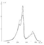
Fig. 1. Absorption spectra of TbIG: solid curve –  ||[110], dashed –
  ||[110],
  ||[001]

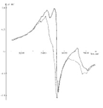
Fig. 2. Frequency dependences of the contribution made to the refractive index by  ^
^ ||[
||[ ]; the optical transition 7F6 – 7F0 of the Tb3+ ion of the TbIG: solid curve –
  ||[110],
  ||[]; dashed –
  ||[110],
  ||[001]
Fig. 1 shows the measured absorption coefficient k’(ħω), in the Voight geometry, of an Tb3Fe5012 a plate cut in the (110) plane, at  ||[110],
  ||[
||[ ], Fig. 2 shows the n’(ħω) spectra calculated on these results with the aid of the Kramers-Kronig relations (n’ is the contribution made to the refractive index by the light absorption by the rare earth (RE) ions). A difference between k’(ħω) and n’(ħω) at two E orientations is evidence that the crystal is optically biaxial. This difference in k’ is a maximum at the frequencies 5450 and 5500 cm-1. It reaches 60-70 %. As seen from Fig. 2, Δn=n’100–n’110 reaches a value 1x10⁻³ and reverses sign several times in the region of the absorption band.
Thus the character of the optical anisotropy of the crystal depends on the relative orientation of the vectors k and I. No such effect had been observed before for Tb3Fe5O12 and for Tb3Ga5O12 in such geometry of the experiment to our knowledge.
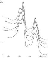
Fig. 3 shows the transmission spectra of linearly polarized light of a terbium gallate. The spectra are obtained in a Voight geometry (the light wave vector k is normal to a surface in which the magnetic field vector H lies). Within the limits of experimental accuracy the spectra for the perpendicular and parallel orientations of the vector E to the direction of H are in agreement. A single-component structure for the transition 7F6 – 7F0 is observed at the 30K temperature and three components at T=100K. At both 30K and 100K for the transition 7F6 – 7F1 two components are observed.
In contrast to the gallate-garnet absorption spectra in the terbium IG, the pattern of the absorption line structure in analogous transitions is complicated, as is seen in Fig.4: broadening of each of the components is observed, and it becomes more difficult to resolve the fine structure of the transitions. As is seen from a comparison, these spectra disclose a substantial optical anisotropy of the magnetic birefringence and dichroism. In formulating this experiment we started from the fact that only the lowest of the thirteen singlets into which the ground multiplet of the terbium ion 7F6 is split by the crystalline field will be populated at low temperatures, while the excited level 7F0 has a singlet structure because the total moment equals zero, and its position will depend relatively weakly on the orientation of I in the crystal. Consequently, the position of the highly energetic components of the absorption line fine structure in the electronic transition 7F6 – 7F0 (and apparently in the transition 7F6 – 7F1) should yield information directly about the anisotropy of the lowest levels of the multiplet 7F6 of the Tb3+ ion.

Fig. 3. Absorption spectra of linearly polarized light of a Tb3Ga5O12: solid curve – T=30 K, dashed – T=100 K
Acknowledgments
The work is executed at support of the Ministry of education and science of the Russian Federation (the project № 2.2527.2011). Investigations were carried out on the equipment of Natural Sciences Research and Education Centre at North-Ossetian State University.


